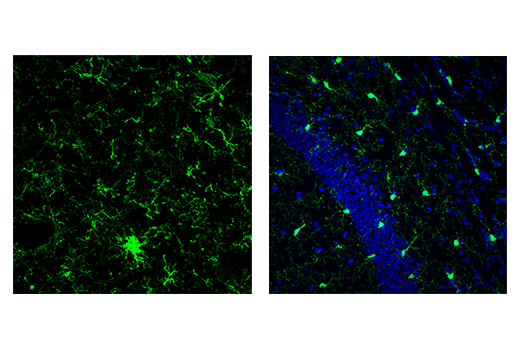
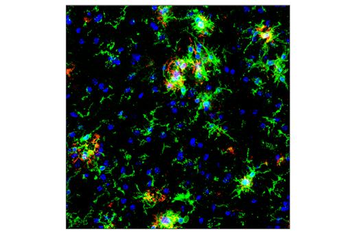
Microglia are a type of glial cell present throughout the brain and spinal cord. As brain-resident macrophages, these cells play an important role as a first line of immune defense in the central nervous system (CNS). Activated microglia can act as antigen presenters and secrete cytokines to trigger further immune responses. With highly sensitive phagocytic processes that extend to multiple locations, they continuously detect and engulf damaged cells, infectious agents, debris, and plaques. Due to their multifaceted ability to monitor and clean up the CNS, these cells are critical for injury repair response and are implicated in neurodegenerative diseases. Microglia are also necessary during brain development as they are critical for dendritic pruning and, in a mature brain, they help maintain a homeostatic environment. Microglia are often identified by observing the expression of cell-specific intracellular proteins, such as Iba1/AIF-1, CD11b, and TMEM119. Click here to see a full list of microglia cell markers. Many of these products are also available conjugated to fluorophores across the spectrum.
Iba1/AIF-1 is an evolutionarily conserved calcium-binding protein. As an F-actin-binding protein, Iba1/AIF-1 may remodel the actin cytoskeleton of microglia. Iba1/AIF-1 is uniquely expressed in cells of monocytic lineage and, therefore, antibodies targeting this protein are widely used to label microglia/macrophages in the brain and other tissue.
The transmembrane protein, Cluster of Differentiation Molecule 11b (CD11b)/Integrin alpha M (ITGAM) consists of α and β heterodimers that are expressed in cells of the innate immune system. Antibodies that detect CD11b/ITGAM are commonly used to identify cells of myeloid lineage, including neutrophils, monocytes, macrophages, and microglia.
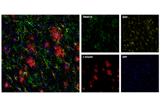
TMEM119 is a transmembrane protein of unknown function that is specifically expressed in almost all microglia. This developmentally regulated protein is not expressed by macrophages or other immune cell types. Antibodies that detect TMEM119 act as specific cell surface markers to visualize microglia.
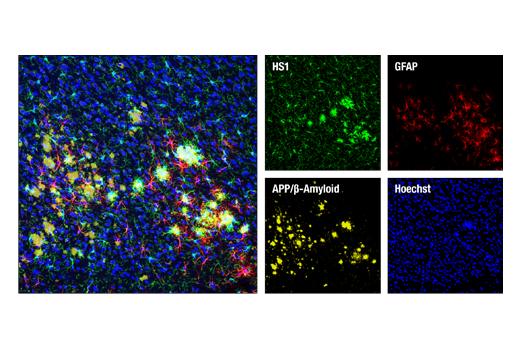
HS1 is a protein kinase substrate that is enriched in cells of hematopoietic origin. In the brain, HS1 is expressed in microglia and monocyte-derived macrophages. HS1 antibodies are useful tools to highlight the cytoplasm of microglia and macrophages.
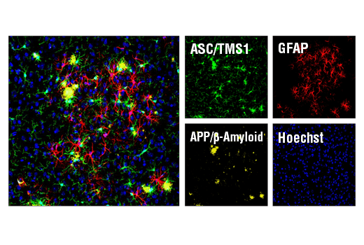
ASC/TMS1 is an adaptor protein, encoded by the PYCARD gene in humans, that is often expressed in the nucleus of monocytes, microglia, and macrophages. This pro-apoptotic protein is a key mediator of the inflammasome that is triggered in response to pathogen infection or tissue damage. During this process, it relocalizes to the perinuclear space, cytoplasm, and endoplasm, as well as the mitochondria where it interacts with Bax to trigger cytochrome c release and cause subsequent apoptosis. ASC/TMS1 is also a crucial component of inflammatory signaling because it activates caspase-1 in response to pro-inflammatory signals.