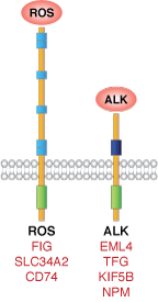
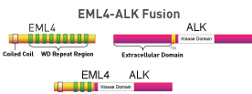
Anaplastic lymphoma kinase (ALK) and c-ros oncogene 1 (ROS1) are related receptor tyrosine kinases (RTKs). Research studies have identified ALK and ROS as mutant C-terminal fusion proteins that are expressed in a wide range of cancers (1-10). Table 1 lists some of the ALK and ROS1 fusion partners that have been identified and the associated cancer types.
| RTKs | Inhibitors | Fusion Partners | Associated Cancers |
|---|---|---|---|
| ALK | Crizotinib5 | EML42,11, TFG1, KIF5B6, NPM7 | NSCLC1,2, ALCL7, CRC11 |
| ROS1 | Crizotinib5 | FIG8, CD749, SLC34A29,11 | Glioblastoma8, Cholangiocarcinoma10, Ovarian cancer3, NSCLC1,4, CRC11 |
Table 1. Summary of ALK and ROS1 fusion partners and associated cancers. Abbreviations: ALCL=Anaplastic large cell lymphoma EML4=Echinoderm microtubule-associated protein-like 4 NPM=Nucleophosmin NSCLC=Non-small cell lung cancer TFG=TRK-fused gene
Cell Signaling Technology recommends two highly sensitive antibodies for detection of ALK and ROS1 full-length proteins and C-terminal fusion proteins.
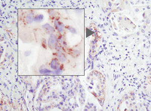
Immunohistochemical analysis of paraffin-embedded human lung carcinoma using ROS1 (D4D6) Rabbit mAb #3287. Note: Staining is of FIG-ROS1 fusion (4).
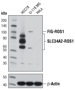
Western blot analysis of extracts from HCC78 (SLC34A2-ROS1), U-118 MG (FIG-ROS1), and HeLa (ROS1 negative) cells using ROS1 (D4D6) Rabbit mAb #3287 (upper) or β-Actin (D6A8) Rabbit mAb #8457 (lower). Note: HCC78 cells express the 85, 70, and 59 kDa forms of the SLC34A2-ROS1 fusion protein (1).
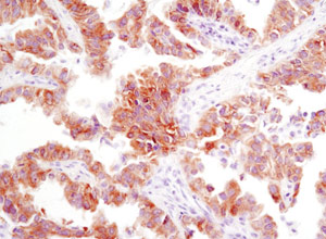
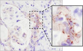
Immunohistochemical analysis of paraffin-embedded human lung carcinoma using ALK (D5F3) XP® Rabbit mAb #3633.
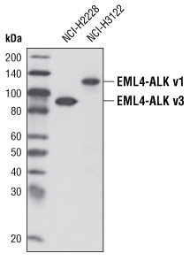
Western blot analysis of extracts from NCI-H2228 and NCI-H3122 cells using ALK (D5F3) XP® Rabbit mAb #3633. Variants denoting fusions of different EML4 exons (v1 or v3) are indicated.
CST performed an unbiased, large-scale survey of tyrosine kinase activity in lung cancer using PTMScan® Technology. This proprietary technology, developed at Cell Signaling Technology(11), uses a CST™ Motif Antibody for immunoaffinity purification of peptides from digested cell extracts combined with LC tandem mass spectrometry to identify and quantify changes in post-translational modifications such as phosphorylation, acetylation, or ubiquitination. For this study, we used a phospho-tyrosine motif antibody to analyze changes in phosphorylation across the proteome in NSCLC cell lines and tissues.
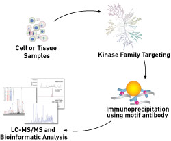
Using PTMScan® Technology, we surveyed the phosphotyrosine status of receptor tyrosine kinases (RTK) and non-receptor tyrosine kinases in 41 NSCLC cell lines and over 150 NSCLC tumors. Over 50 tyrosine kinases and more than 2,500 downstream substrates that play roles in NSCLC growth and progression were identified. Two very exciting findings from this study were the identification of novel anaplastic lymphoma kinase (ALK) and c-ros oncogene 1 (ROS1) C-terminal fusion proteins in some NSCLC cell lines and tumors.
Rabbit monoclonal antibodies specific to ALK and ROS1 [ALK (D5F3) XP® Rabbit mAb #3633; ROS1 (D4D6) Rabbit mAb #3287] were developed to detect both full-length and C-terminal fusion proteins. These antibodies have been validated for IHC and can detect ALK and ROS1 fusion protein expression in NSCLC sample(12, 4).
IHC analysis of paraffin-embedded human lung carcinoma using ROS1 (D4D6) Rabbit mAb #3287. Note: Staining is of FIG-ROS1 fusion protein(4).
A partnership with Pfizer, Inc., creator of the ALK inhibitor crizotinib, and Ventana Medical Systems, Inc., a leader in companion diagnostic testing, was formed to develop the use of ALK (D5F3) XP® Rabbit mAb in an automated diagnostic IHC screening assay to detect ALK fusion proteins in NSCLC patient samples.
Patient samples that stain positive for ALK expression may be candidates for crizotinib, which was approved for use in the U.S. in August 2011.
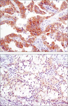
IHC analysis of paraffin-embedded human lung carcinoma with high (upper) and low levels (lower) of ALK expression using ALK (D5F3) XP® Rabbit mAb #3633.
CST scientists are highlighted in bold.