View Featured Offers >>
48669
CAR (G4S Linker) Cell Enrichment Kit
Cellular Assay Kits
Assay Kit

CAR (G4S Linker) Cell Enrichment Kit #48669

Citations (0)
Flow cytometric analysis of live cells in the input (left) and post-enrichment (right) for enrichment performed using biotinylated G4S Linker (E7O2V) Rabbit mAb. Input consists of CD4+/CD8+ human T cells containing a mixture of non-transduced cells and cells transduced with an scFv-based Anti-CD20 (Leu16) CAR containing a G4S linker. The post-enrichment sample shows a nearly pure population of cells expressing the CAR on the cell surface. Anti-rabbit IgG (H+L), F(ab')2 Fragment (Alexa Fluor® 647 Conjugate) #4414 was used as a secondary antibody to detect the biotinylated antibody.
Flow cytometric analysis of live cells in the input (left) and post-enrichment (right) for enrichment performed using biotinylated G4S Linker (E7O2V) Rabbit mAb. Input contains a mixture of WT Jurkat cells and Jurkat cells engineered to stably express an scFv-based Anti-CD19 (FMC63) CAR containing a G4S linker. Tag Blue fluorescent protein (TagBFP) is co-expressed with the CAR. The post-enrichment sample shows a nearly pure population of cells expressing the CAR on the cell surface. Anti-rabbit IgG (H+L), F(ab')2 Fragment (Alexa Fluor® 647 Conjugate) #4414 was used as a secondary antibody to detect the biotinylated antibody. Engineered cell line was provided by the Lohmueller Lab, University of Pittsburgh.
To Purchase # 48669
Cat. # Size Qty. Price
48669S
1 Kit

Product Includes Quantity (with Count)
Lyophilized DNase I 3 x 1 ea
Releasable Streptavidin Beads 1 x 5 ml
DNase I Reconstitution Buffer 1 x 2 ml
Biotinylated G4S Linker (E7O2V) Rabbit mAb 2 x 1250 µl

Protocol

PRINT

View >Collapse >

Cell Enrichment Kit Protocol

NOTE: Kit capacity: ~2 x 10⁹ total input cells

A. Solutions and Reagents

Supplied Reagents
  • Releasable Streptavidin Beads (1 bottle, 5 mL #27119)
  • Lyophilized DNase I (3 vials, > 15,000 units each #91019)
  • DNase I Reconstitution Buffer (1 bottle, 2 mL #47776)
  • Biotinylated Rabbit mAb (specific to kit)
Additional Reagents (Not Supplied)

NOTE: Refer to Table 1 for recommended tube sizes and volumes.

  • Magnet appropriate for tube size
  • Mixer allowing tilting and rotation of tubes
  • Phosphate Buffered Saline with BSA (PBS-B): Ca2+ and Mg2+ free PBS supplemented with 0.1% bovine serum albumin (BSA), pH 7.4
  • Phosphate Buffered Saline with BSA and EDTA (PBS-BE): Ca2+ and Mg2+ free PBS with 0.1% BSA (or 0.1% human serum albumin or fetal calf serum) and 2 mM EDTA (or 0.6% sodium citrate)
  • Cell Elution Buffer: RPMI 1640 medium with 1% fetal calf serum (FCS), 1 mM CaCl2 and 5 mM MgCl2, pH 7.0–7.4
General Considerations:
  • Protocol volumes are suitable for enrichment of 1 x 10⁷ total input cells but are directly scalable up to 2 x 10⁸ cells. For higher or lower total cell input count, refer to Table 1.
  • When enriching for cells expressing targets with high variability of expression, titration of biotinylated mAb and releasable streptavidin beads is recommended to optimize purity and yield.
  • Keep all buffers cold unless otherwise specified.
  • Avoid air bubbles (foaming) during pipetting.

B. Reagent Preparation

NOTE: For optimal DNase I activity, ensure that the Cell Elution Buffer pH is 7.0-7.4.

NOTE: Reconstitute vials of enzyme as needed.

  1. Wash Releasable Streptavidin Beads (#27119):
    1. Resuspend stock vial of beads via vortexing >30 sec or tilt and rotate for 5 min.
    2. Transfer the recommended volume of beads for the number of purifications being performed to a tube (refer to step 5 in Table 1).
    3. Add the same volume, or at least 1 mL, of PBS-B and mix.
    4. Place the tube in a magnet for 1 min, discard the supernatant.
    5. Remove the tube from the magnet and resuspend washed beads in the same volume of PBS-B as the initial volume of beads.
  2. Reconstitute DNase I:
    1. To each vial of Lyophilized DNase I #91019 (as needed), add 300 μL of DNase I Reconstitution Buffer #47776.
    2. Dissolve the enzyme gently. Vigorous mixing (e.g., pipetting/vortexing too hard) will destroy its activity.
    3. Aliquot reconstituted DNase I into suitable portions.
    4. Store at –20°C.

      NOTE: Thaw immediately before use and keep on ice once thawed. Thawed reconstituted DNase I can be refrozen once.

  3. Warm appropriate volume of Cell Elution Buffer for the number of purifications being performed to 37°C (refer to step 13 in Table 1).

C. Cell Preparation

NOTE: Blood and serum may contain soluble factors (e.g., antibodies or cell surface antigens) which can interfere with the cell isolation protocol. Washing the cells once may reduce this interference.

NOTE: Optimal centrifugation conditions will vary depending upon cell type and reagent volume. Generally, 180-350 x g for 5-10 min will be sufficient to pellet the cells.

  1. Collect cells of interest and pellet by centrifugation.
  2. Resuspend cells at 1 x 10⁷ cells/mL in PBS-BE at 2-8°C.

D. Cell Enrichment

NOTE: For steps requiring incubations with gentle tilting and rotation, do not perform end-over-end mixing if the volume is small relative to the tube size. Tilt and rotate so the cells and beads are kept in the bottom of the tube.

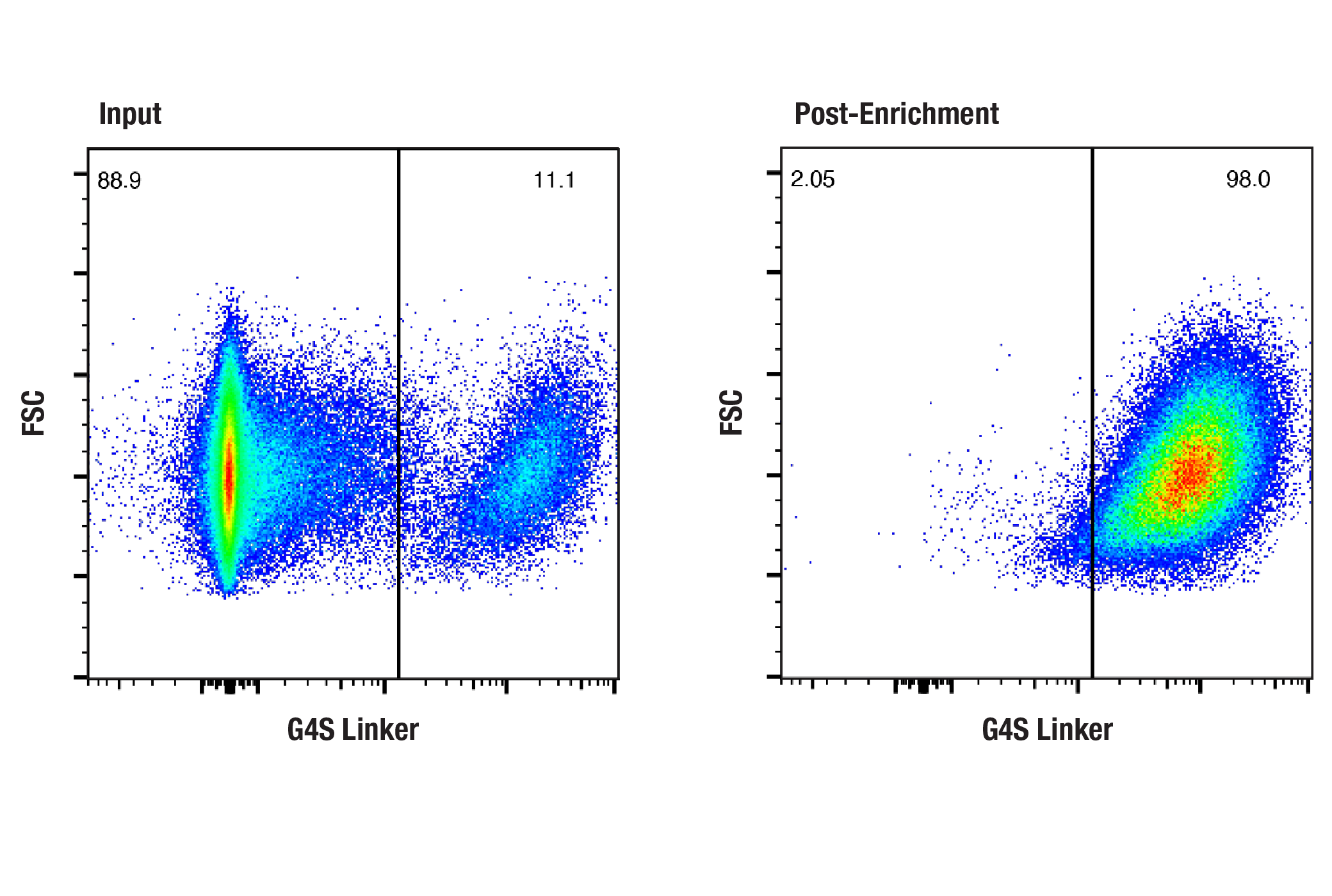
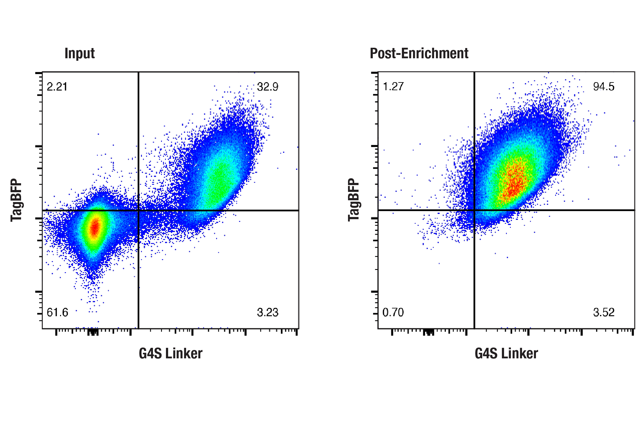
NOTE: If desired, a small sample of resuspended cells from step 4 can be stained with a fluorochrome-conjugated secondary antibody (e.g., Anti-rabbit IgG (H+L), F(ab')2 Fragment (Alexa Fluor® 647 Conjugate) #4414) to determine the percentage of target expressing cells in the input prior to enrichment.

NOTE: Purity of elution fraction can be assessed by flow cytometry using a fluorochrome-conjugated secondary antibody (e.g., Anti-rabbit IgG (H+L), F(ab')2 Fragment (Alexa Fluor® 647 Conjugate) #4414).

  1. Add 12.5 µL biotinylated antibody to 1 mL cell suspension, mix gently.
  2. Incubate for 10 min at 2-8°C.
  3. Wash cells by adding 2 mL PBS-BE, then pellet by centrifugation. Discard the supernatant.
  4. Resuspend cells at 1 x 10⁷ cells/mL in PBS-BE.
  5. Resuspend pre-washed beads and add 25 µL to cells.
  6. Incubate 20 min at 2-8°C with gentle tilting and rotation.
  7. Optional: Add 1 mL PBS-BE to limit trapping of unbound cells.
  8. Place the tube in a magnet for 2 min.
  9. While the tube is still in the magnet, remove and discard the supernatant.
  10. Remove the tube from the magnet and add 1 mL PBS-B, pipet 2-3 times or vortex 2-3 sec, replace tube in magnet for 2 min.
  11. Repeat steps 9 and 10 at least twice to wash the cells. This step is critical to obtain high purity of CAR positive cells.
  12. While the tube is still in the magnet, remove and discard the supernatant.
  13. Resuspend bead-bound cells in 400 µL pre-warmed (37°C) Cell Elution Buffer.
  14. Add 4 µL reconstituted DNase I.
  15. Incubate for 15 min at room temperature with gentle tilting and rotation.

    NOTE: Before transferring released cells to new tube in step 17, pre-coat collection tubes with Cell Elution Buffer for at least 5 min.

  16. Pipet thoroughly at least 5-10 times to maximize cell release (avoid foaming).
  17. Place in magnet for 2 min, then transfer the supernatant with released cells into a tube pre-coated with Cell Elution Buffer.
  18. Remove tube from magnet, resuspend bead fraction in 400 µL Cell Elution Buffer, repeat steps 16 and 17 once to collect residual cells.

Table 1. Recommended Volumes for Different Cell Numbers

Cell Enrichment Step Step Description Volumes Per 1 x 10⁷ Total Input Cells Volumes Per 2 x 10⁸ Total Input Cells
  Recommended tube size 5 mL 50 mL
  Recommended magnet e.g., Invitrogen DynaMag-5 e.g., Invitrogen DynaMag-50
1* Biotinylated mAb 12.5 µL 250 µL
1* Cell volume 1 mL 20 mL
3** Wash cells (PBS-BE) 2 mL 40 mL
4 Resuspend cells (PBS-BE) 1 mL 20 mL
5*** Add beads 25 µL 500 µL
7 Optional: Increase volume (PBS-BE) 1 mL 20 mL
10-11** Wash cells (PBS-B) 3 x 1 mL 3 x 20 mL
13 Resuspend cells (Cell Elution Buffer) 400 µL 8 mL
14 Release cells (reconstituted DNase I) 4 µL 80 µL
18 Collect residual cells (Cell Elution Buffer) 400 µL 8 mL

*If total cell input count is lower than 1 x 10⁷ cells, adjust biotinylated antibody volume and keep cell density at 1 x 10⁷ cells/mL. Wash volumes can be kept the same as for 1 x 10⁷ cells.

**Adjust buffer volumes to fit the tube you are using.

***If the target expressing cell population is high (e.g., > 2.5 x 10⁶ target cells/mL), increase the amount of beads (maximum double the amount).

Protocol Id: 3164

Product Description

The CAR (G4S Linker) Cell Enrichment Kit is designed to positively select cells that are engineered to express a G4S linker-containing single-chain variable fragment (scFv)-based chimeric antigen receptor (CAR) on the cell surface. Using an indirect magnetic bead-based immunoaffinity enrichment protocol, this kit yields highly pure and viable CAR positive cells that are bead-free. Within a heterogeneous population of cells, desired CAR expressing cells are labeled with a biotinylated rabbit monoclonal antibody targeting the G4S linker. Releasable Streptavidin Beads are then incubated with the cell mixture. Bead-bound cells are isolated without columns using a magnet (not included), and then beads are removed from cells using DNase I. Following release from beads, the positively selected fraction of CAR positive cells containing surface-bound biotinylated antibody-streptavidin complexes are available for expansion or use in downstream applications such as flow cytometry and gene expression analyses.

Bead Specifications:
Releasable Streptavidin Beads are paramagnetic polystyrene beads (4.5 μm diameter) coupled to streptavidin via a DNA linker susceptible to cleavage by DNase I. The beads provided in this kit are not recommended for the isolation of phagocytic cells as the beads are likely to be engulfed by these cells.

Specificity / Sensitivity

Biotinylated G4S Linker (E7O2V) Rabbit mAb recognizes exogenously expressed levels of scFv-based CARs containing a G4S linker.

Species Reactivity:

All Species Expected

Background

Magnetic bead-based immunoaffinity cell enrichment is a gentle purification method that can be leveraged, in part, to facilitate a robust interrogation of the biology of immune cells that are engineered to express CARs. For example, isolation of rare subsets of CAR positive cells from a complex, heterogeneous population of cells presents the opportunity for extensive downstream analyses using single-cell omics assays. In the context of CAR cell engineering, magnetic bead-based immunoaffinity cell enrichment can also be useful in scenarios when delivery of the CAR transgene is inefficient, resulting in a small population of cells expressing the CAR transgene.

Limited Uses

Except as otherwise expressly agreed in a writing signed by a legally authorized representative of CST, the following terms apply to Products provided by CST, its affiliates or its distributors. Any Customer's terms and conditions that are in addition to, or different from, those contained herein, unless separately accepted in writing by a legally authorized representative of CST, are rejected and are of no force or effect.

Products are labeled with For Research Use Only or a similar labeling statement and have not been approved, cleared, or licensed by the FDA or other regulatory foreign or domestic entity, for any purpose. Customer shall not use any Product for any diagnostic or therapeutic purpose, or otherwise in any manner that conflicts with its labeling statement. Products sold or licensed by CST are provided for Customer as the end-user and solely for research and development uses. Any use of Product for diagnostic, prophylactic or therapeutic purposes, or any purchase of Product for resale (alone or as a component) or other commercial purpose, requires a separate license from CST. Customer shall (a) not sell, license, loan, donate or otherwise transfer or make available any Product to any third party, whether alone or in combination with other materials, or use the Products to manufacture any commercial products, (b) not copy, modify, reverse engineer, decompile, disassemble or otherwise attempt to discover the underlying structure or technology of the Products, or use the Products for the purpose of developing any products or services that would compete with CST products or services, (c) not alter or remove from the Products any trademarks, trade names, logos, patent or copyright notices or markings, (d) use the Products solely in accordance with CST Product Terms of Sale and any applicable documentation, and (e) comply with any license, terms of service or similar agreement with respect to any third party products or services used by Customer in connection with the Products.

For Research Use Only. Not for Use in Diagnostic Procedures.
Cell Signaling Technology is a trademark of Cell Signaling Technology, Inc.
All other trademarks are the property of their respective owners. Visit our Trademark Information page.