| Cat. # | Size | Qty. | Price |
|---|---|---|---|
| 9961T | 1 Kit (6 x 20 microliters) |
|
| Product Includes | Quantity | Applications | Reactivity | MW(kDa) | Isotype |
|---|---|---|---|---|---|
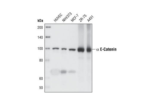
| α-E-Catenin (23B2) Rabbit mAb 3240 | 20 µl |
|
H M Mk | 100 | Rabbit IgG |
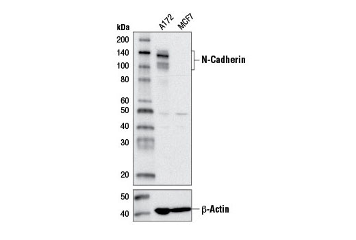
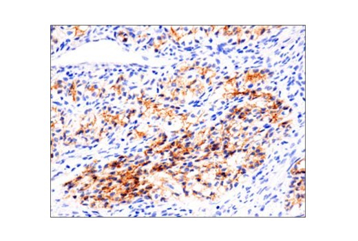
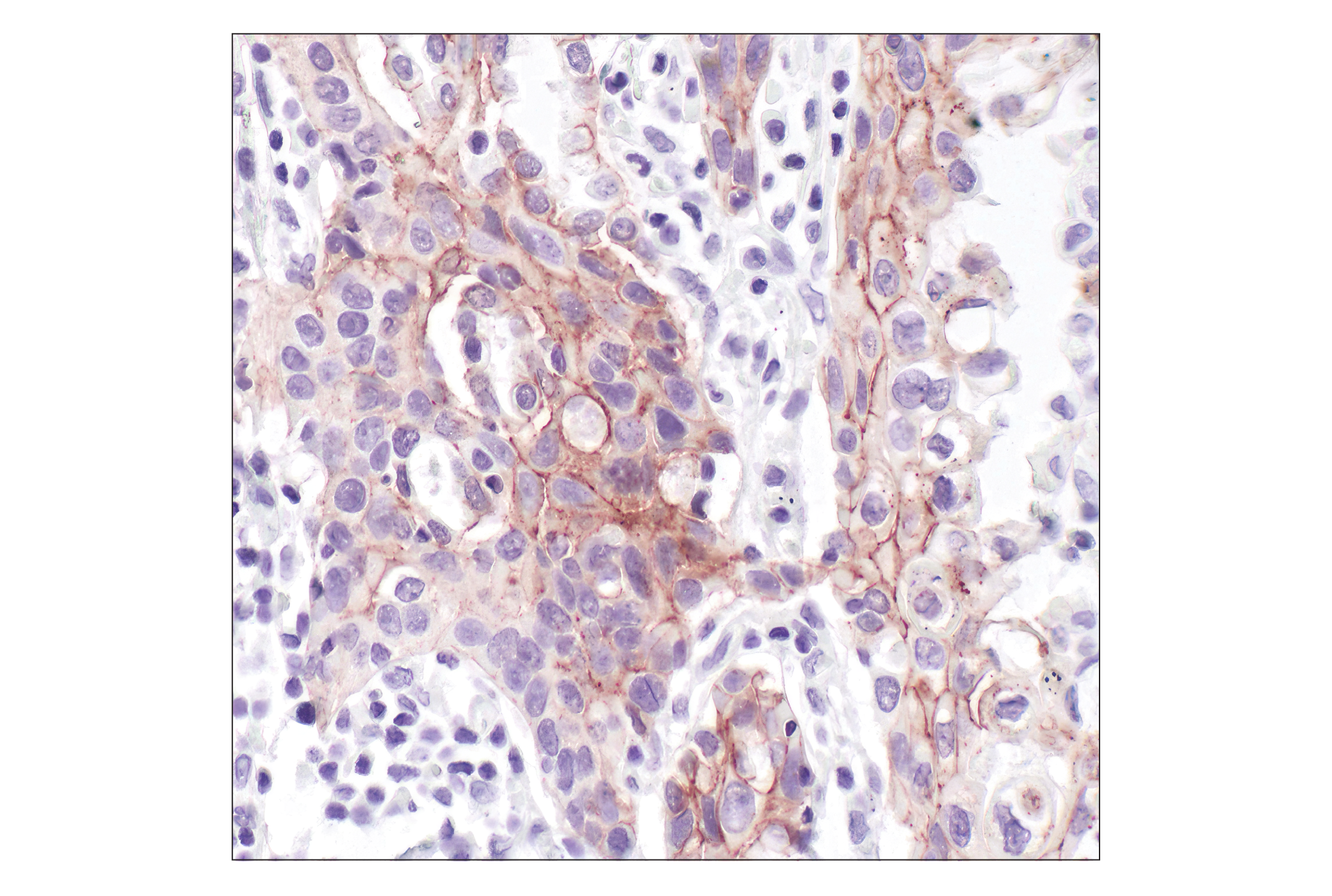
| N-Cadherin (D4R1H) XP® Rabbit mAb 13116 | 20 µl |
|
H M | 140 | Rabbit IgG |
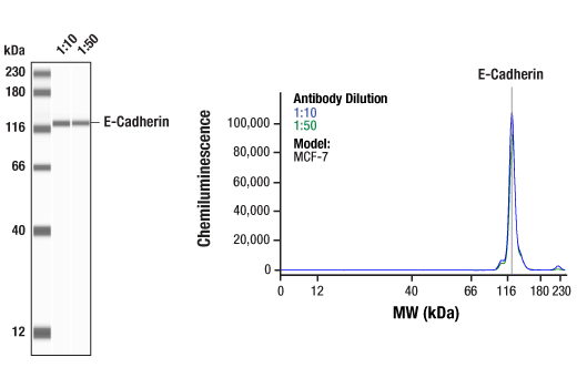
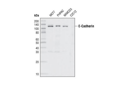
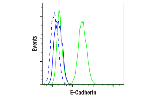
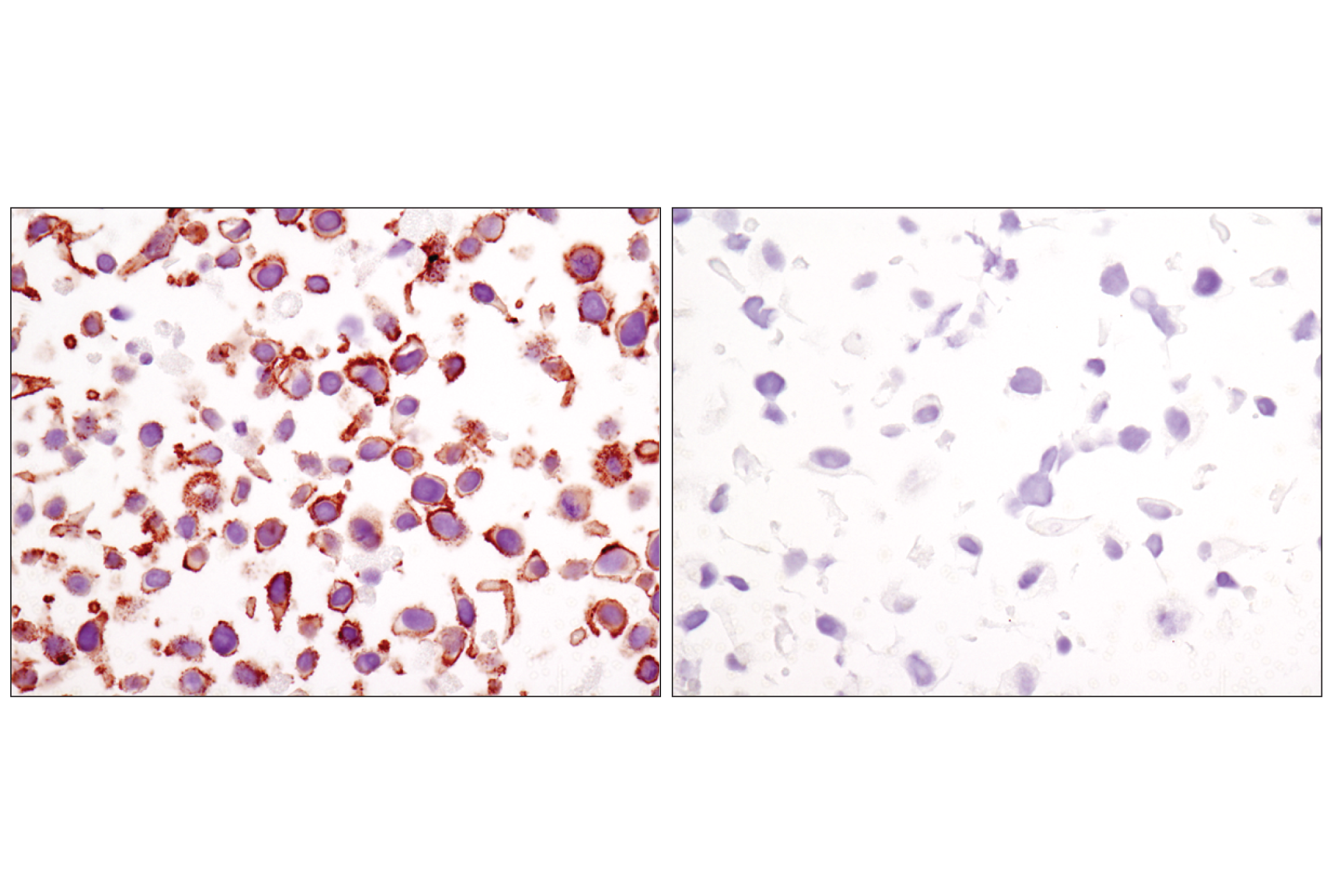
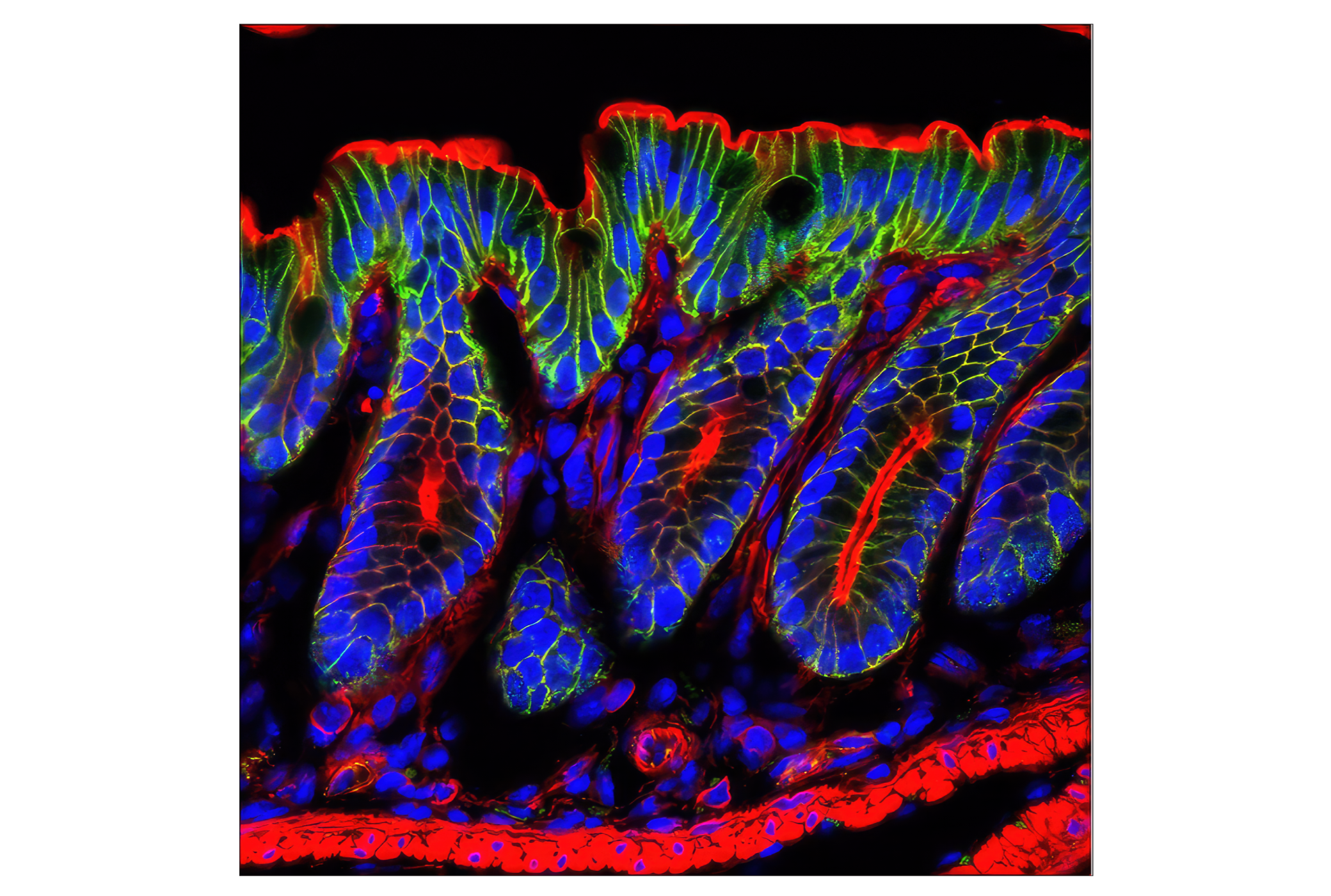
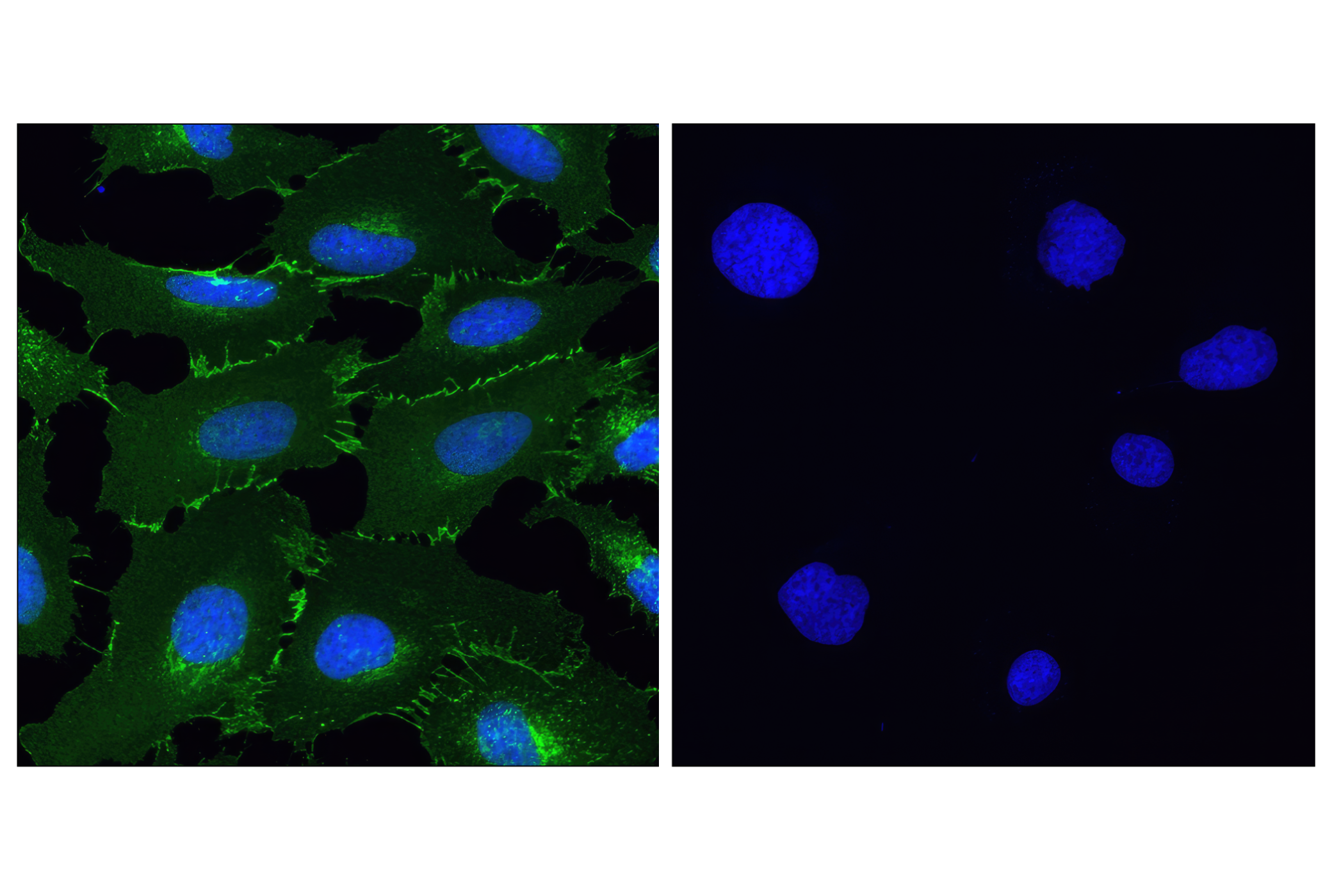
| E-Cadherin (24E10) Rabbit mAb 3195 | 20 µl |
|
H M | 135 | Rabbit IgG |
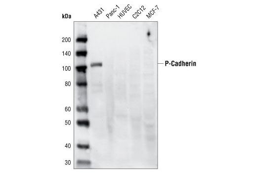
| P-Cadherin (C13F9) Rabbit mAb 2189 | 20 µl |
|
H | 120 | Rabbit IgG |
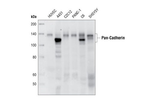
| Pan-Cadherin (28E12) Rabbit mAb 4073 | 20 µl |
|
H M R | 130-150 | Rabbit IgG |
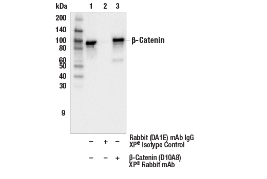
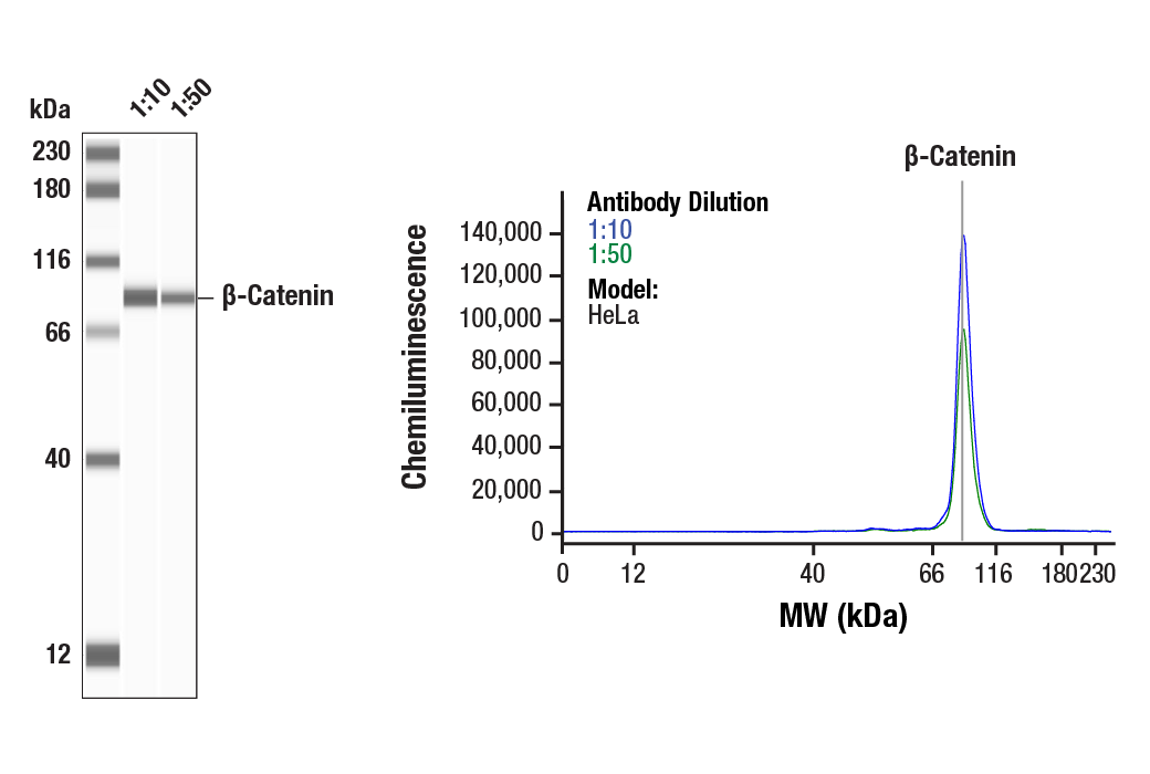
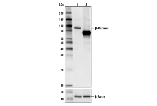
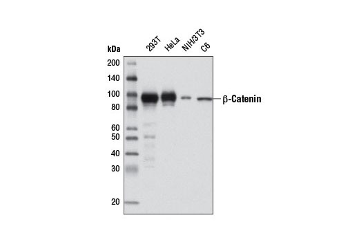
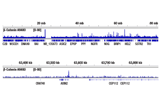
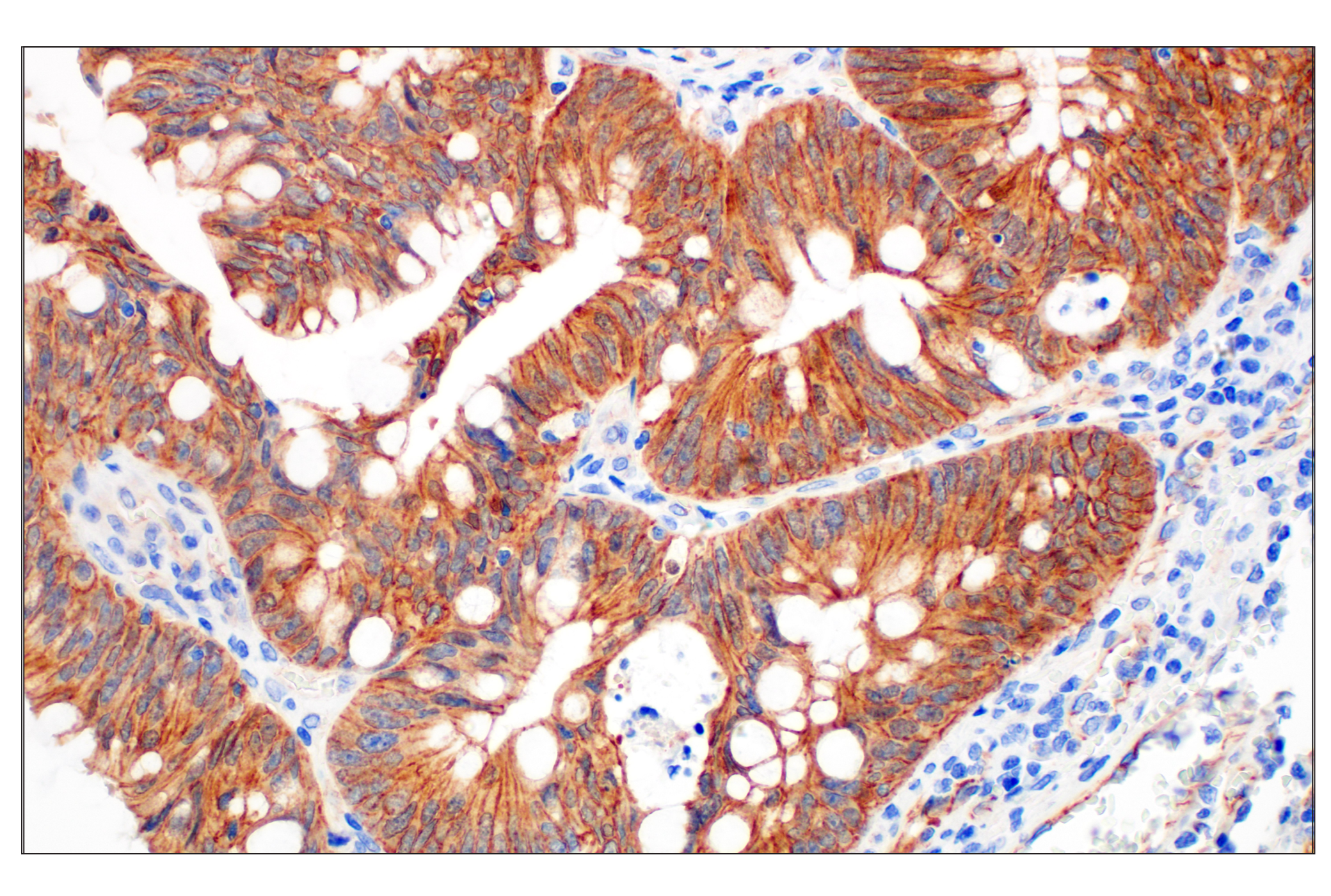
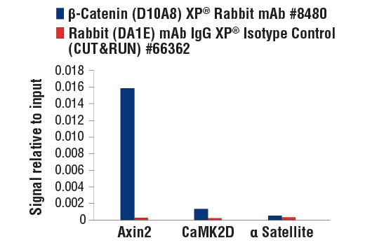
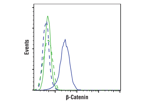
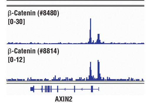
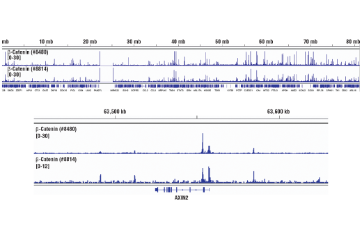
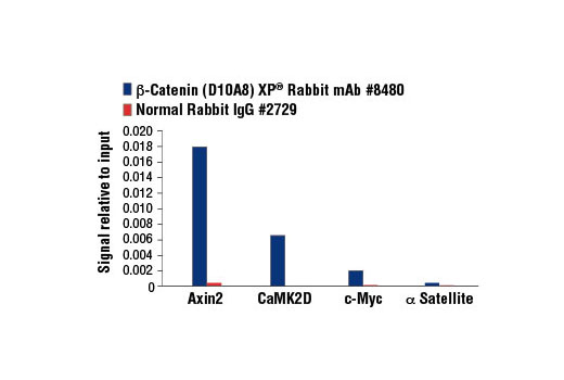
| β-Catenin (D10A8) XP® Rabbit mAb 8480 | 20 µl |
|
H M R Mk | 92 | Rabbit IgG |
| Anti-rabbit IgG, HRP-linked Antibody 7074 | 100 µl |
|
Rab | Goat |
Product Information
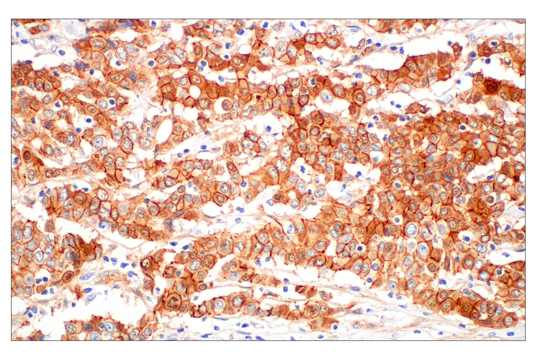
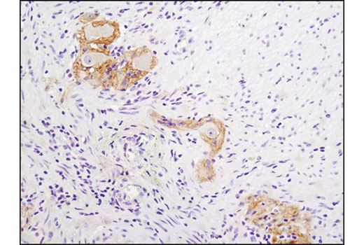
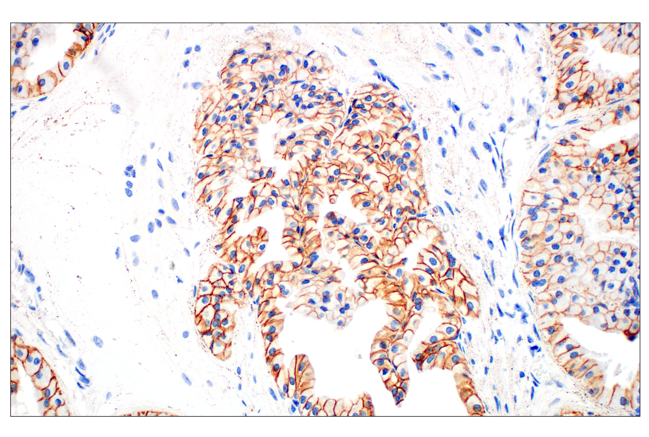
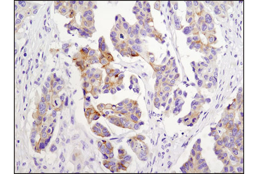
Monoclonal antibody is produced by immunizing animals with a synthetic peptide corresponding to the amino-terminal sequence of human α-E-catenin, residues surrounding Arg526 of human N-cadherin protein, residues near the carboxy terminus of human P-cadherin, residues surrounding 780 of human E-cadherin, residues surrounding Pro714 of human ß-catenin protein, and a synthetic peptide corresponding to a conserved region of human N-, R-, E- and P-Cadherin.
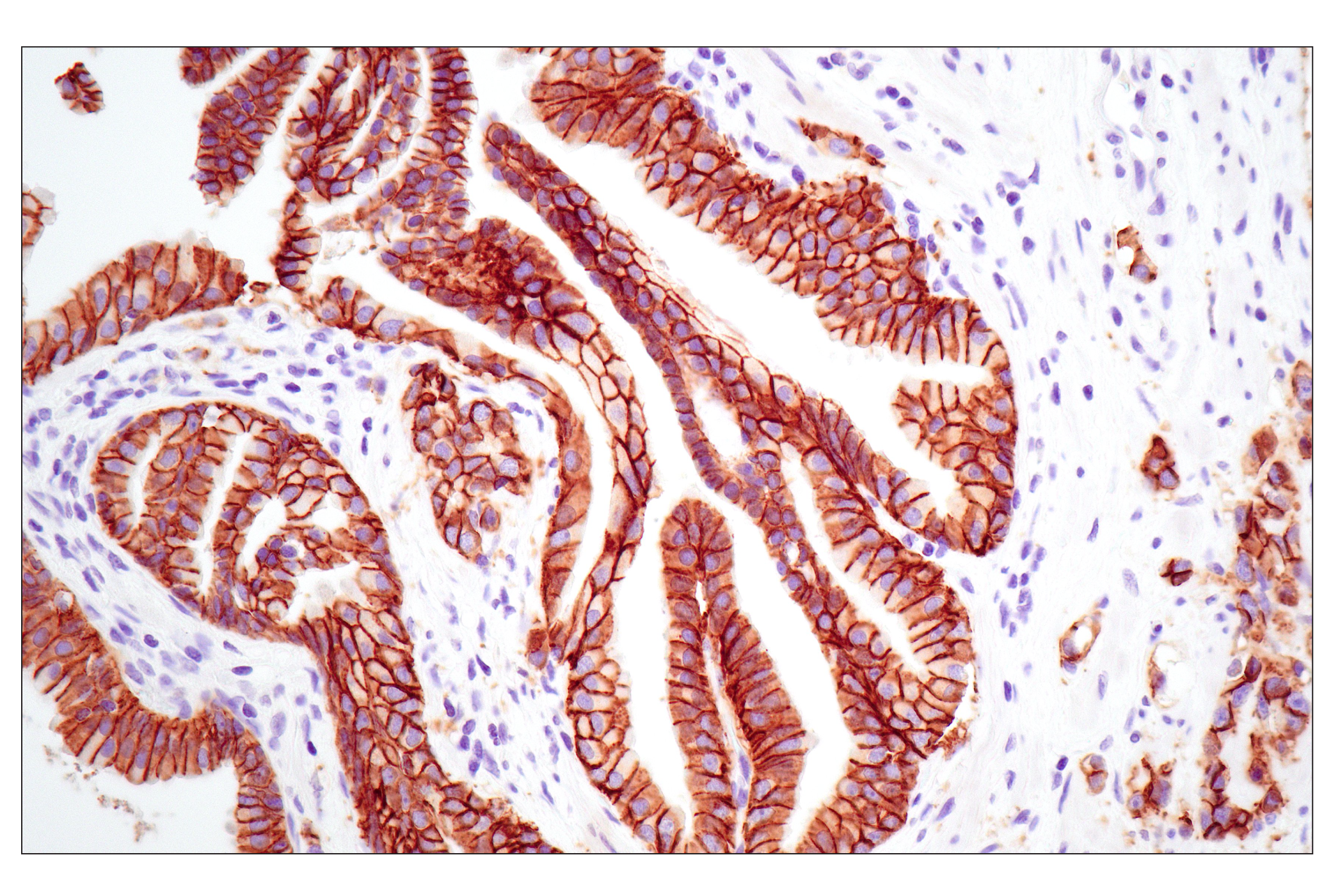
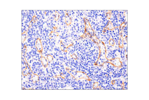
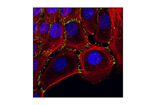
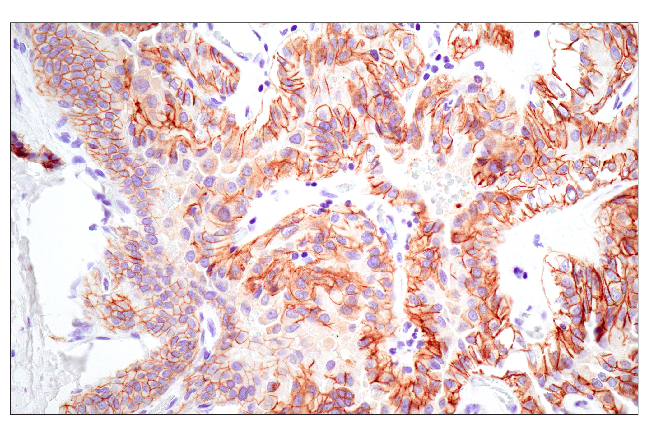
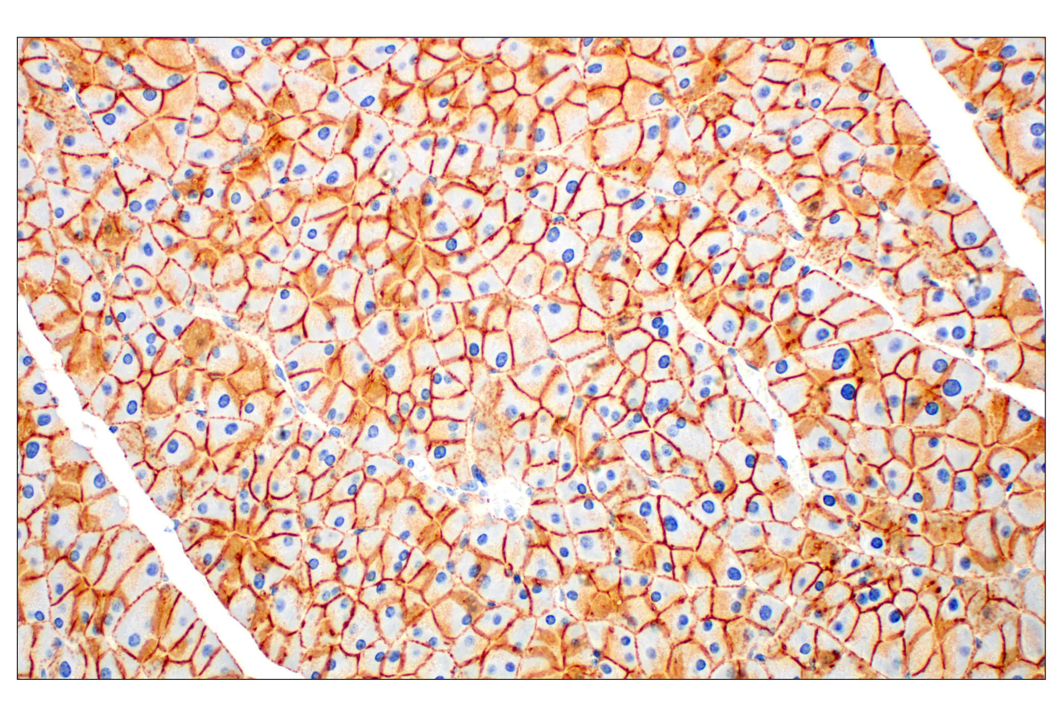
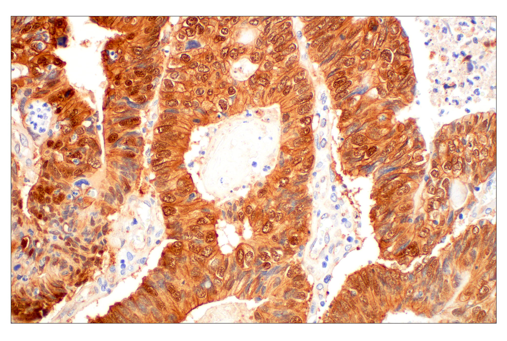
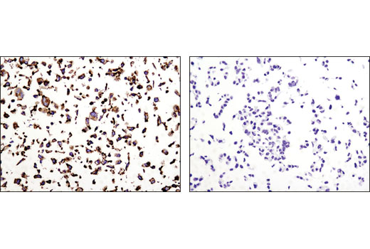
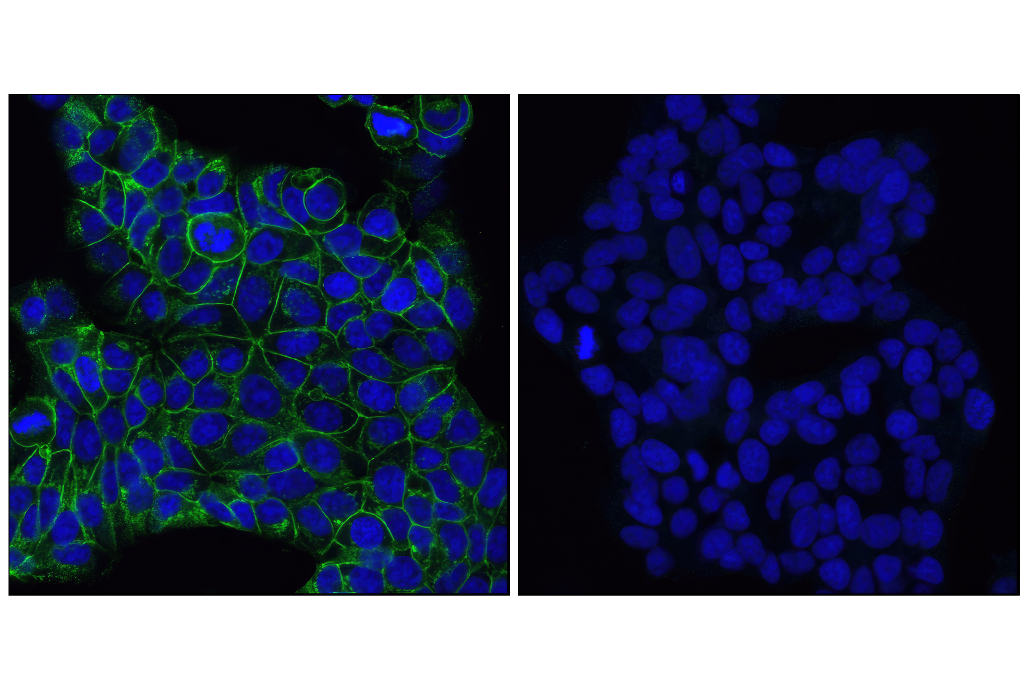
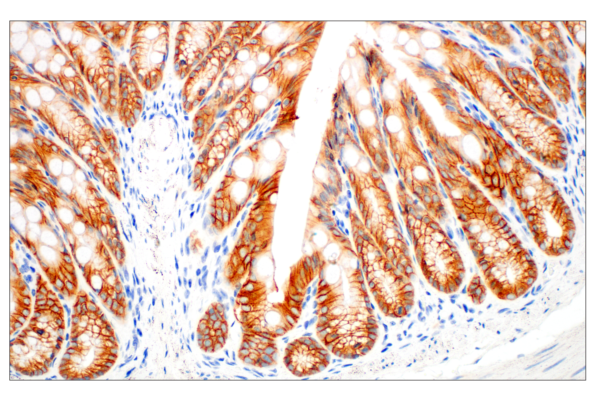
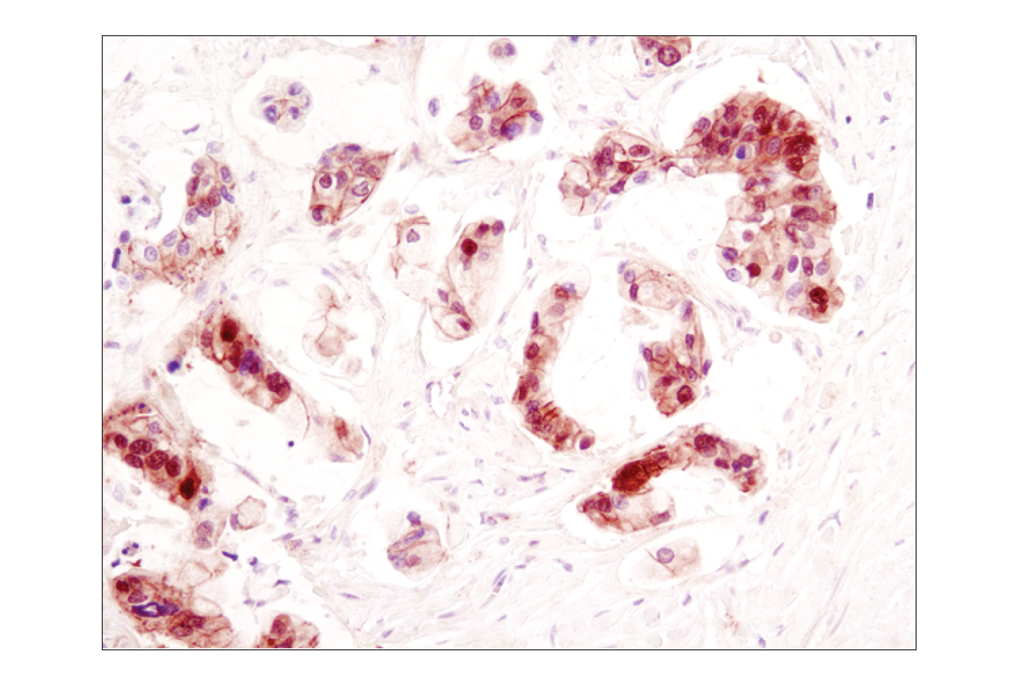
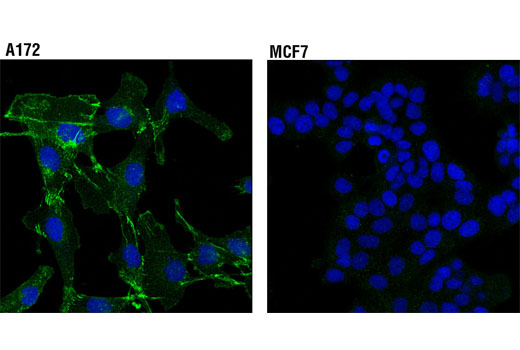
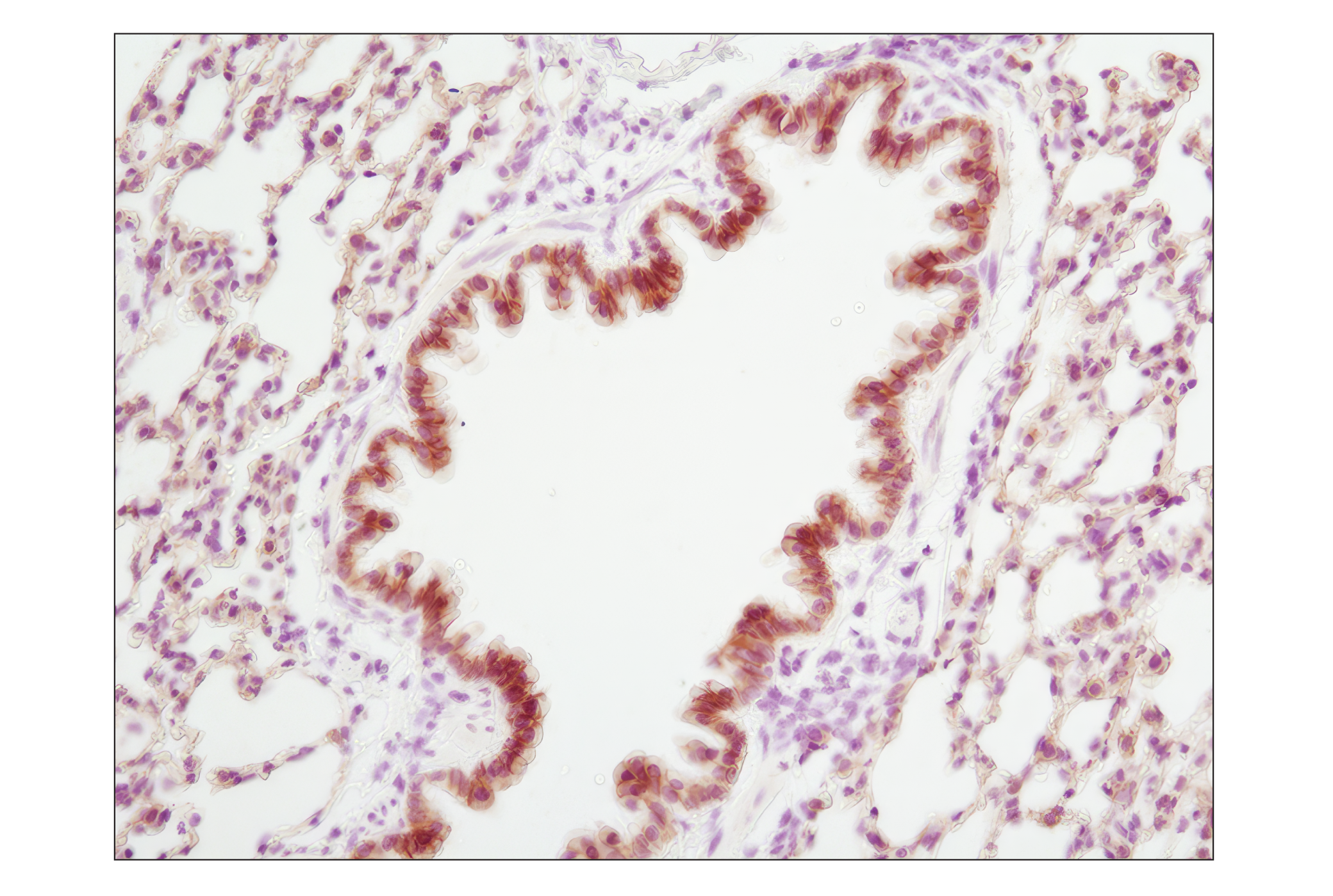
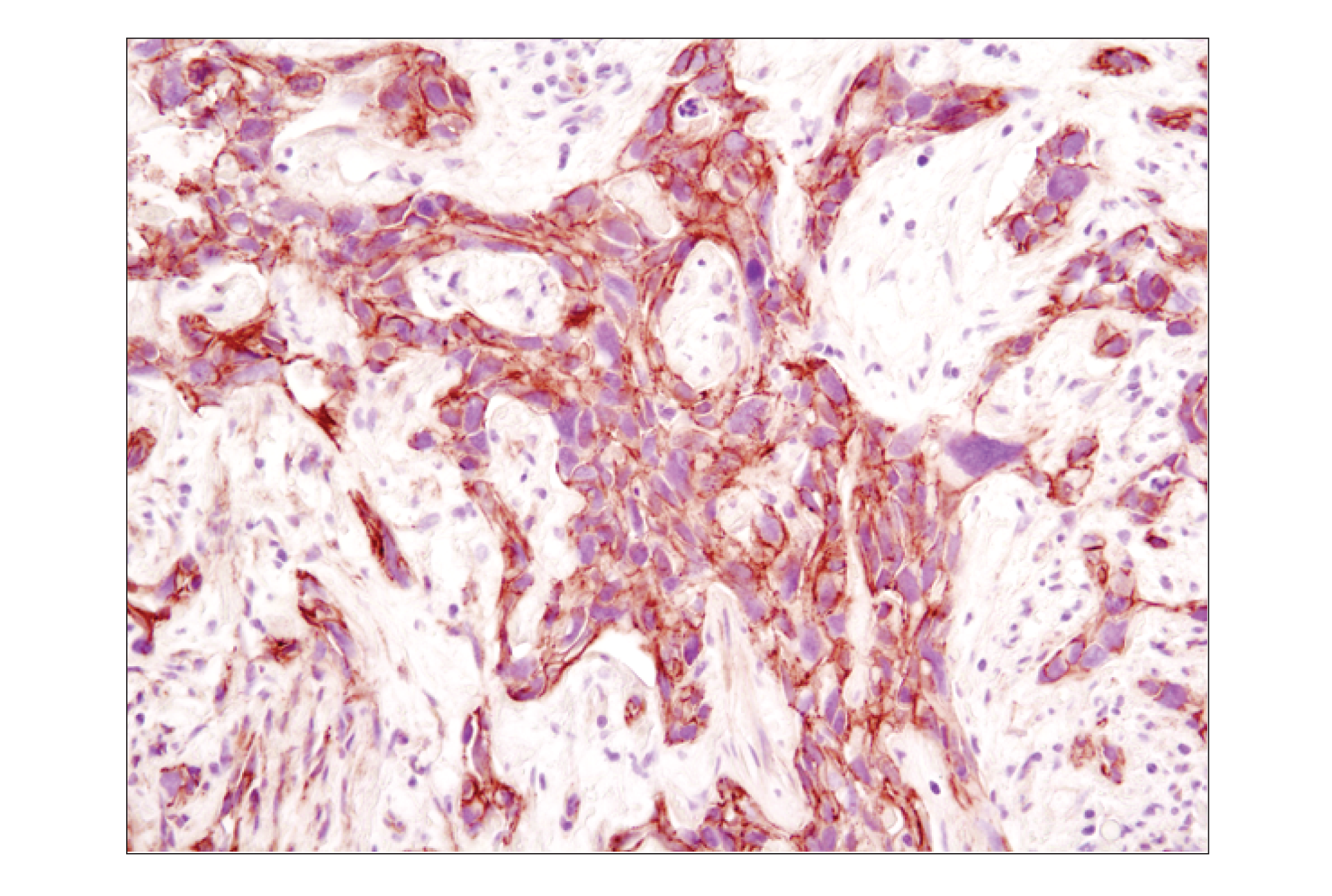
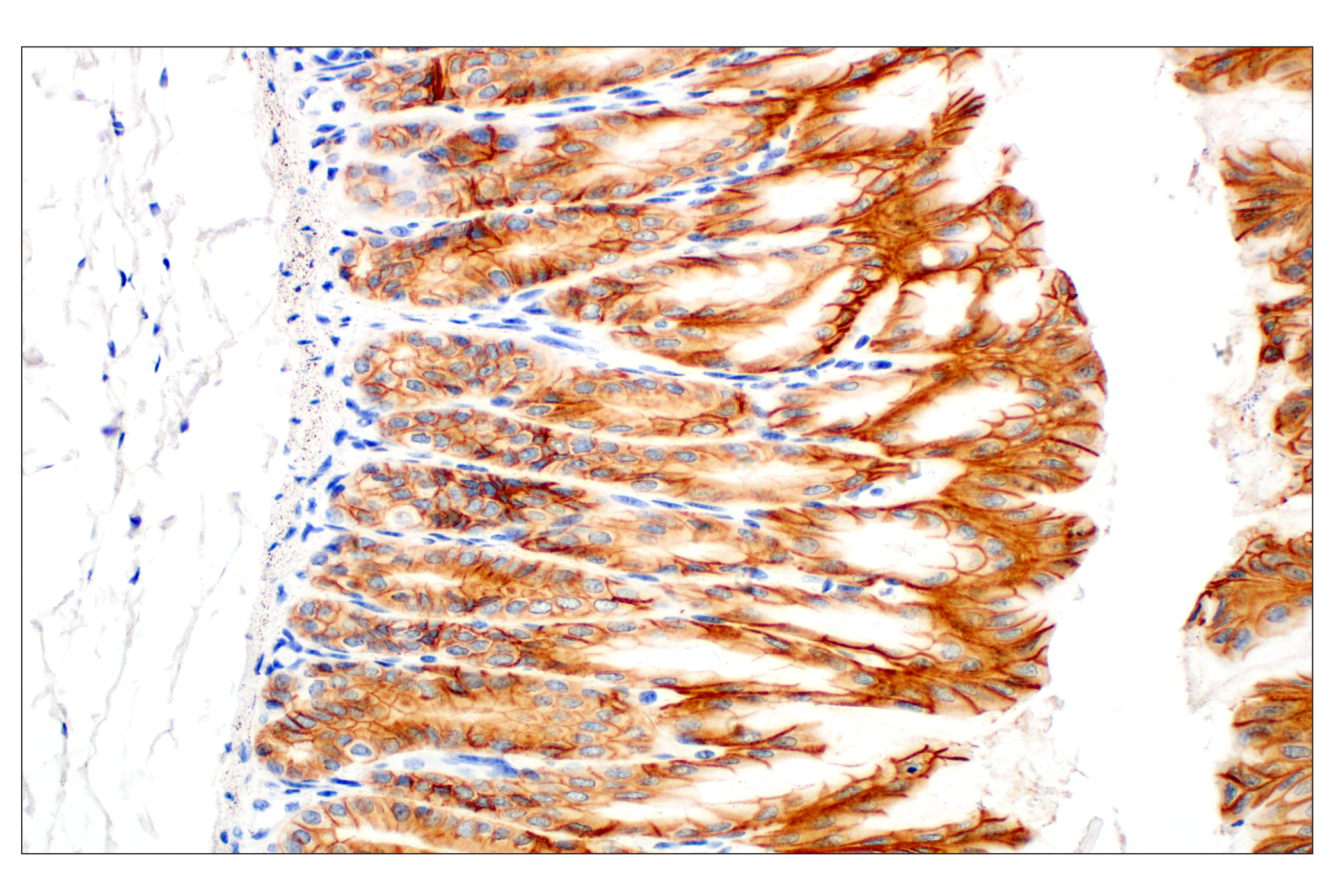
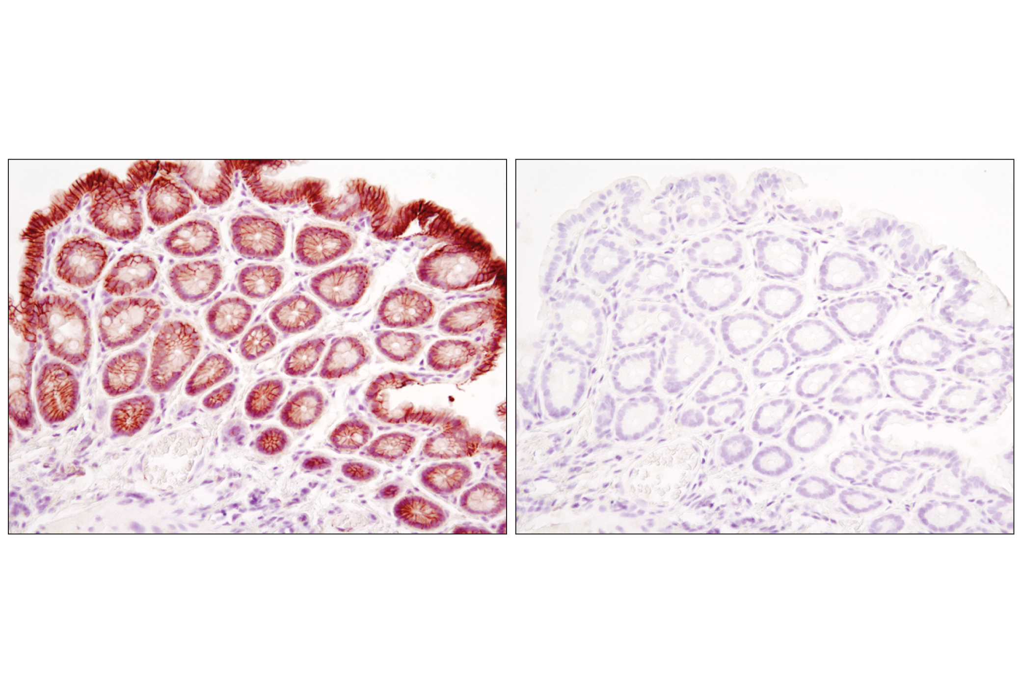
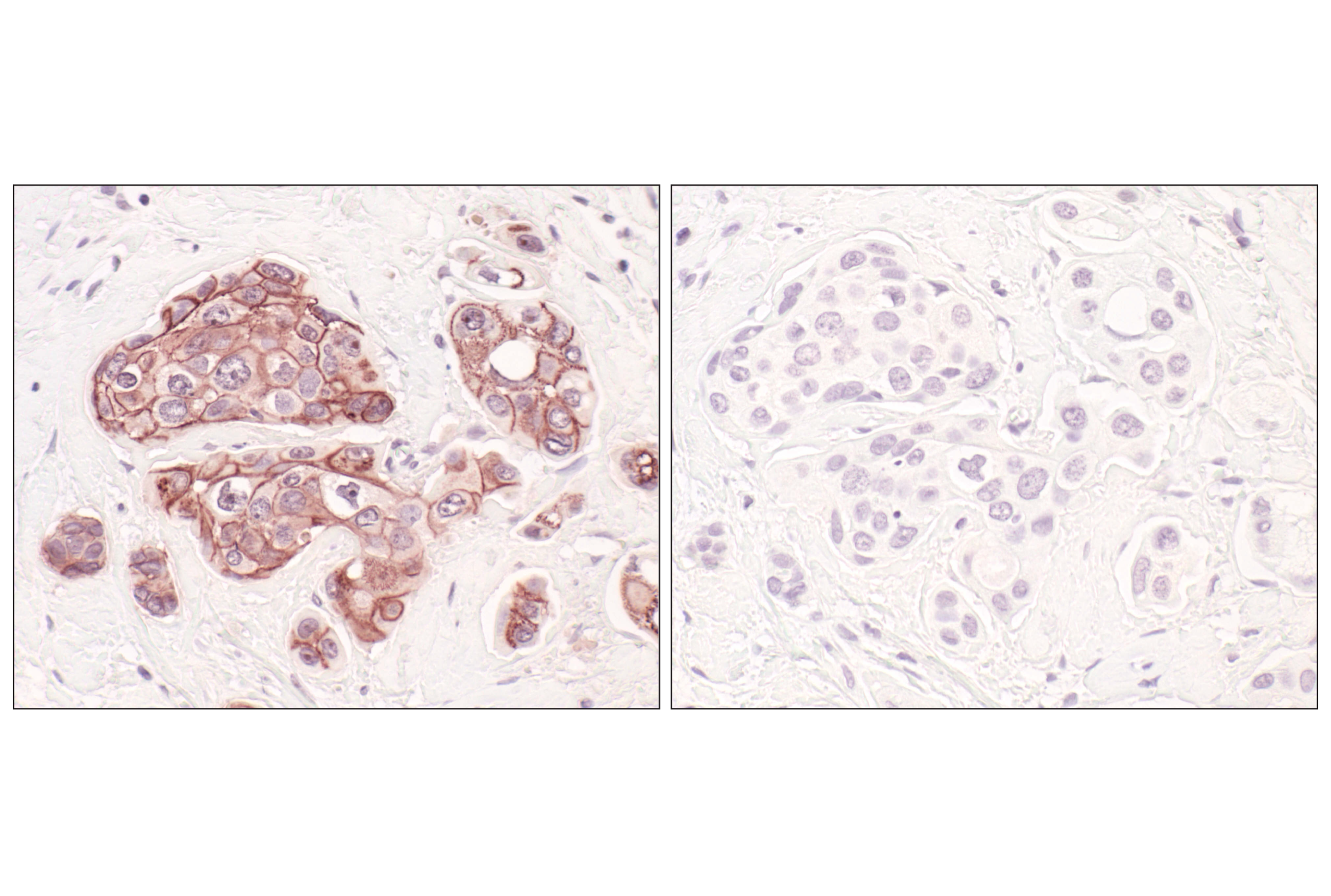
Adherens junctions are dynamic structures that form cell-cell contacts and are important in development, differentiation, tissue integrity, morphology and cell polarity. They are composed of cadherins that are transmembrane proteins that bind cadherins on adjacent cells in a calcium dependent manner. On the cytoplasmic side of adherens junctions, the cadherins associate with β-catenin, γ-catenin and p120 catenin (δ). β-catenin and γ-catenin associate with α-catenin, which links the cadherin-catenin complex to the actin cytoskeleton (1,2). Recent studies indicate that cancer cells exhibit increased N-cadherin and diminished E-cadherin expression. E-cadherin is considered a suppressor of invasive cancer cell growth and this change in cadherin expression associated with cancer progression is termed the “cadherin switch”. β-catenin is one of the key downstream effectors in the Wnt signaling pathway and has been implicated in early embryonic development and tumorigenesis (3-5).
Explore pathways related to this product.
STRING - Known and Predicted Protein-Protein Interactions.
Except as otherwise expressly agreed in a writing signed by a legally authorized representative of CST, the following terms apply to Products provided by CST, its affiliates or its distributors. Any Customer's terms and conditions that are in addition to, or different from, those contained herein, unless separately accepted in writing by a legally authorized representative of CST, are rejected and are of no force or effect.
Products are labeled with For Research Use Only or a similar labeling statement and have not been approved, cleared, or licensed by the FDA or other regulatory foreign or domestic entity, for any purpose. Customer shall not use any Product for any diagnostic or therapeutic purpose, or otherwise in any manner that conflicts with its labeling statement. Products sold or licensed by CST are provided for Customer as the end-user and solely for research and development uses. Any use of Product for diagnostic, prophylactic or therapeutic purposes, or any purchase of Product for resale (alone or as a component) or other commercial purpose, requires a separate license from CST. Customer shall (a) not sell, license, loan, donate or otherwise transfer or make available any Product to any third party, whether alone or in combination with other materials, or use the Products to manufacture any commercial products, (b) not copy, modify, reverse engineer, decompile, disassemble or otherwise attempt to discover the underlying structure or technology of the Products, or use the Products for the purpose of developing any products or services that would compete with CST products or services, (c) not alter or remove from the Products any trademarks, trade names, logos, patent or copyright notices or markings, (d) use the Products solely in accordance with CST Product Terms of Sale and any applicable documentation, and (e) comply with any license, terms of service or similar agreement with respect to any third party products or services used by Customer in connection with the Products.