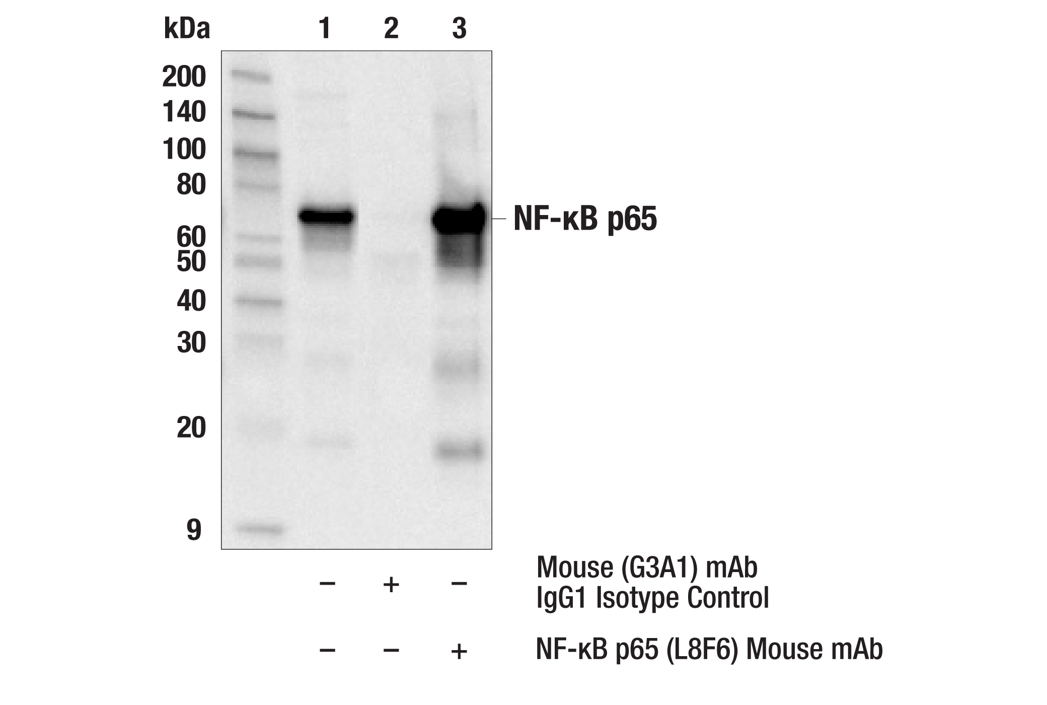
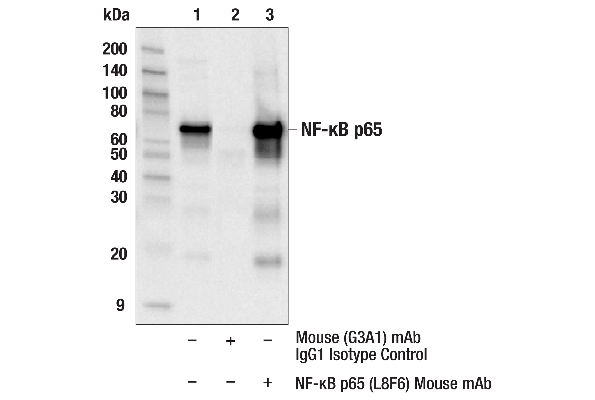
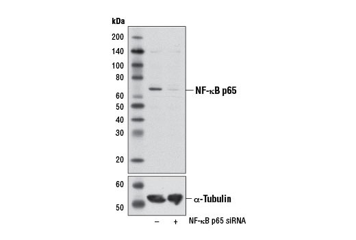
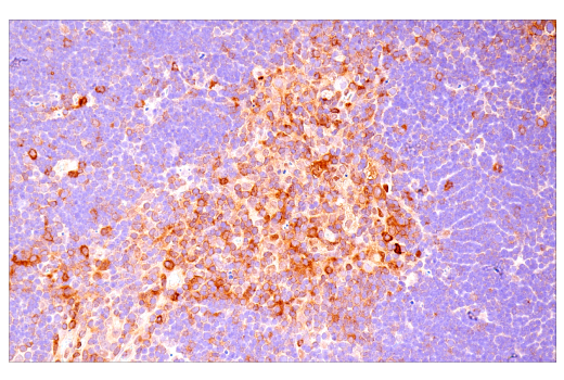
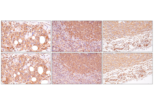
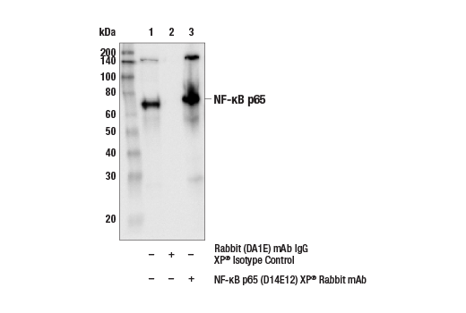
Immunoprecipitation of NF-kB p65 from HeLa cell extracts. Lane 1 is 10% input, lane 2 is precipitated with Mouse (G3A1) mAb IgG1 Isotype Control #5415, and lane 3 is NF-κB p65 (L8F6) Mouse mAb, #6956. Western blot was performed using NF-κB p65 (D14E12) XP® Rabbit mAb, #8242.
| Cat. # | Size | Qty. | Price |
|---|---|---|---|
| 4766T | 1 Kit (8 x 20 microliters) |
|
| Product Includes | Quantity | Applications | Reactivity | MW(kDa) | Isotype |
|---|---|---|---|---|---|
| NF-κB p65 (L8F6) Mouse mAb 6956 | 20 µl |
|
H M R Hm Mk Mi B Dg Pg | 65 | Mouse IgG2b |
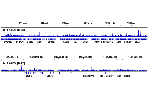
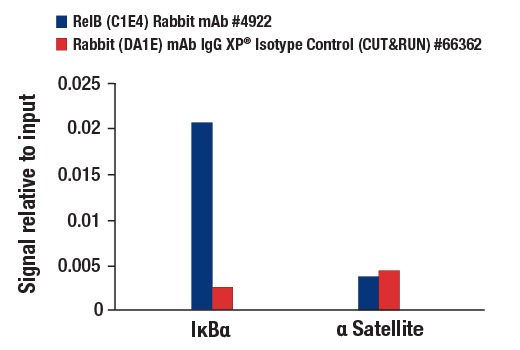
| RelB (C1E4) Rabbit mAb 4922 | 20 µl |
|
H M R Mk | 70 | Rabbit IgG |
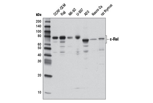
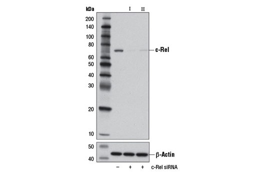
| c-Rel (D4Y6M) Rabbit mAb 12707 | 20 µl |
|
H M R | 68-78 | Rabbit IgG |
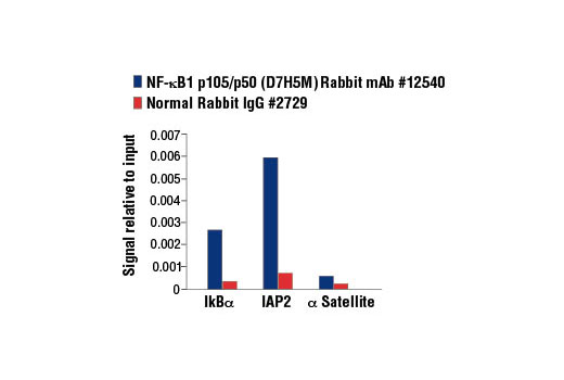
| NF-κB1 p105/p50 (D7H5M) Rabbit mAb 12540 | 20 µl |
|
H M | 50 Active form. 120 Precursor | Rabbit IgG |
| NF-κB1 p105 Antibody 4717 | 20 µl |
|
H M R Mk Mi B Pg | 120 | Rabbit |
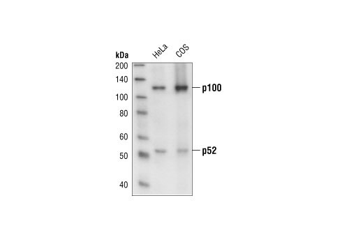
| NF-κB2 p100/p52 Antibody 4882 | 20 µl |
|
H M R Mk | 52 (mature). 120 (precursor). | Rabbit |
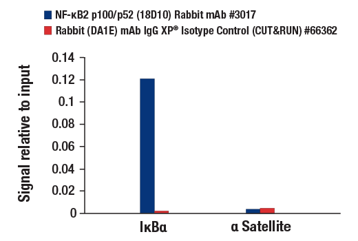
| NF-κB2 p100/p52 (18D10) Rabbit mAb 3017 | 20 µl |
|
H Mk | 52 active form. 120 precursor. | Rabbit IgG |
| NF-κB p65 (D14E12) XP® Rabbit mAb 8242 | 20 µl |
|
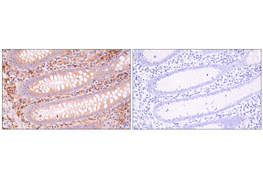
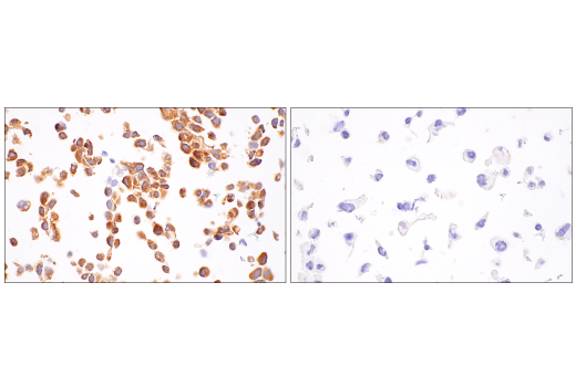
H M R Hm Mk Dg | 65 | Rabbit IgG |
| Anti-rabbit IgG, HRP-linked Antibody 7074 | 100 µl |
|
Goat | ||
| Anti-mouse IgG, HRP-linked Antibody 7076 | 100 µl |
|
M | Horse |
Product Information
Monoclonal antibodies are produced by immunizing animals with synthetic peptides corresponding to amino acid residues near the carboxy terminus of human NF-κB p65, surrounding Ser424 of human RelB, surrounding Leu65 of human c-Rel protein, surrounding Gly415 of human NF-κB p105/p50 protein, surrounding Glu498 of human NF-κB p65/RelA protein, and near the amino terminus of human NF-κB2 p100/p52. Polyclonal antibodies are produced by immunizing animals with synthetic peptides corresponding to amino acid residues at the the carboxy terminus of human NF-κB1 p105 and near the amino terminus of human NF-κB2 p100/p52. Polyclonal antibodies are purified by Protein A and peptide affinity chromatography.
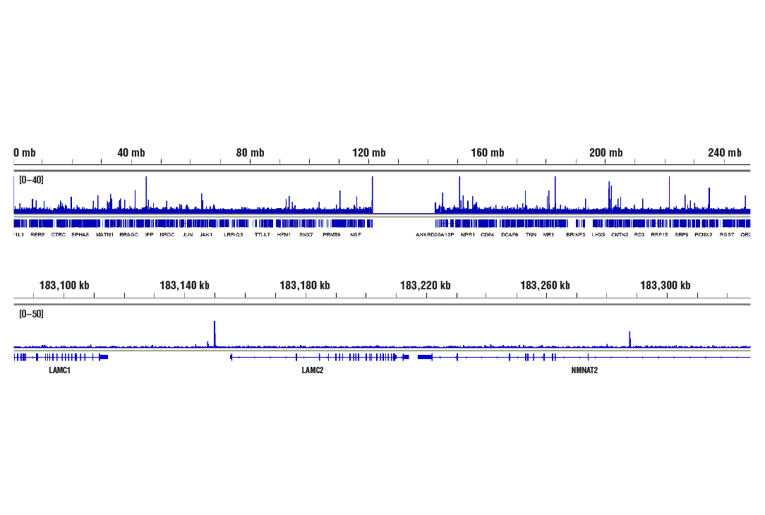
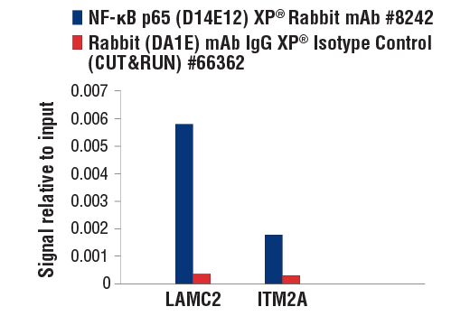
Transcription factors of the nuclear factor κB (NF-κB)/Rel family play a pivotal role in inflammatory and immune responses (1,2). There are five family members in mammals: RelA, c-Rel, RelB, NF-κB1 (p105/p50), and NF-κB2 (p100/p52). Both p105 and p100 are proteolytically processed by the proteasome to produce p50 and p52, respectively. Rel proteins bind p50 and p52 to form dimeric complexes that bind DNA and regulate transcription. In unstimulated cells, NF-κB is sequestered in the cytoplasm by IκB inhibitory proteins (3-5). NF-κB-activating agents can induce the phosphorylation of IκB proteins, targeting them for rapid degradation through the ubiquitin-proteasome pathway and releasing NF-κB to enter the nucleus where it regulates gene expression (6-8). NIK and IKKα (IKK1) regulate the phosphorylation and processing of NF-κB2 (p100) to produce p52, which translocates to the nucleus (9-11).
Except as otherwise expressly agreed in a writing signed by a legally authorized representative of CST, the following terms apply to Products provided by CST, its affiliates or its distributors. Any Customer's terms and conditions that are in addition to, or different from, those contained herein, unless separately accepted in writing by a legally authorized representative of CST, are rejected and are of no force or effect.
Products are labeled with For Research Use Only or a similar labeling statement and have not been approved, cleared, or licensed by the FDA or other regulatory foreign or domestic entity, for any purpose. Customer shall not use any Product for any diagnostic or therapeutic purpose, or otherwise in any manner that conflicts with its labeling statement. Products sold or licensed by CST are provided for Customer as the end-user and solely for research and development uses. Any use of Product for diagnostic, prophylactic or therapeutic purposes, or any purchase of Product for resale (alone or as a component) or other commercial purpose, requires a separate license from CST. Customer shall (a) not sell, license, loan, donate or otherwise transfer or make available any Product to any third party, whether alone or in combination with other materials, or use the Products to manufacture any commercial products, (b) not copy, modify, reverse engineer, decompile, disassemble or otherwise attempt to discover the underlying structure or technology of the Products, or use the Products for the purpose of developing any products or services that would compete with CST products or services, (c) not alter or remove from the Products any trademarks, trade names, logos, patent or copyright notices or markings, (d) use the Products solely in accordance with CST Product Terms of Sale and any applicable documentation, and (e) comply with any license, terms of service or similar agreement with respect to any third party products or services used by Customer in connection with the Products.